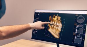
Cone-beam CT is an exciting development in dental and maxillofacial imaging.
This unique Specialist Consultant taught course offers a three day interactive educational experience focusing on the theory behind the safe operation and referral for CBCT (Level 1) and image reporting (Level 2) of both 2D and 3D imaging, primarily for smaller volume CBCT.
Course description
This three-day course builds from the first presentation. Full attendance is required to allow certification.
- Days 1 and 2 cover Level 1 and 2A course material (this allows didactic, group work and interactive workshop elements throughout).
- Day 3 called Level 2B covers essential material not possible to cover in the initial 2 day session.
A learner / delegate-centred approach is adopted throughout, allowing delegates to understand the theory behind the safe operation and referral for CBCT (IRMER Referrer role).
An holistic approach to Image reporting (IRMER Operator Reporter role) covering 2D and 3D imaging primarily for smaller volume CBCT is offered.
Course lead

The course is run by Dr Neil Heath, a Consultant Dental and Maxillofacial Radiologist at Newcastle Dental Hospital.
An experienced educator in CBCT working in NHS, academic and industry sectors, Dr Heath is passionate about delivering relevant enjoyable courses, to like-minded colleagues.
Learning outcomes
The course is delivered in two sessions – initial 2 days followed by a separate final day a few weeks after the first 2 day session to allow reflection and utilisation of new skills.
At the end of this course delegates will be able to:
- Make safe CBCT referrals with a sound understanding of the Physics / Radiation dosages involved and the patient factors to consider before utilising this 3D imaging modality
- Appreciate when 2D and 3D CBCT imaging are best used and how to access guidelines in this regard
- Recapitulate knowledge of 2D dental imaging and learn the anatomical patterns associated with CBCT 3D presentations to know what ‘normal’ looks like
- Appreciate the Radiation safety (IRMER and IRR legislation) surrounding the use of CBCT
- Operate / ‘Drive‘ CBCT viewing software – utilise MPR screens to interrogate the full FOV
- Safely analyse and construct a smaller Vol CBCT report using ‘protocols’ (utilising taught higher order thinking tools ) in a systematic fashion and know when to ask for help
- Analyse examples of concerning dental/maxillofacial disease presentations and know how best to refer on urgent cases. Recognise bone diseases and especially cancers on imaging
- Analyse relevant 2D and 3D image data and construct a recordable imaging report/assessment
- Begin to understand how image reporting errors / imaging pathway adverse events can occur and appreciate how SOPs, Bias and Human factors can influence this process.
Audience
- Dentists (hospital and high street – primary care and specialist), medically qualified specialists involved in imaging the maxillofacial regions.
- Dental Therapists, Dental Nurses or Practice Managers may be interested in attending day 2 (CBCT level 1 material) covered on that day on the course.
Criteria
In order to book on this course you will need:
- GDC / GMC registration (HCPC registration if a Radiographer)
- Those who have a level 1 CBCT certification already may be excused the day 2 element although further cases are reviewed in the workshop on this day
- Those who have already been certified as Level 1 and 2 CBCT on other national courses may wish to use day 3 (CBCT 2B element) as a top up for the CPD in this area